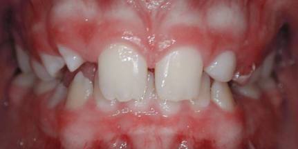
Adolescent Treatment Examples

Adolescent Class I, Crowded

Class-I-crowded-1
The most common malocclusion we treat is the Class Ⅰ crowded bite relationship.

Class-I-crowded-2
The Class Ⅰ notation indicates the position of the first molars in that the upper first molar is slightly behind the lower first molar.

Class-I-crowded-3
This patient had four first premolars removed to eliminate the crowding and allow alignment of the teeth.

Class-I-crowded-4
Finished front photo

Class-I-crowded-5
inished side photo showing closure of all spaces.

Adolescent Class II, Div 1

Class-II-div-1-Ext-U4s-1
A patient with a Class Ⅱ div 1 malocclusion.

Class-II-div-1-Ext-U4s-2
Notice the Class Ⅱ molar (upper molar ahead of the lower).

Class-II-div-1-Ext-U4s-3
This patient was missing the two lower permanent second premolars. The two baby teeth remaining have not erupted as the other teeth have. In this patient these two lower baby teeth were removed and the upper first premolars removed to allow the upper front teeth to be moved back to meet the lower front teeth.

Class-II-div-1-Ext-U4s-4
Finished treatment photo.

Class-II-div-1-Ext-U4s-5
Finished Treatment photo from the side.

Adolescent Class II, Div 1- Example 2

Cl-II-div-1-ext-u4s-les-1
Another common malocclusion seen is the Class Ⅱ patient where the lower teeth are situated behind the upper teeth.

Cl-II-div-1-ext-u4s-les-2
The upper teeth stick out past the lower. Many people call this an “overbite”.

Cl-II-div-1-ext-u4s-les-3
In this patient Dr. Griffies decided to remove two upper first premolars to allow the front teeth to be moved back to meet the lower front teeth.

Cl-II-div-1-ext-u4s-les-4
Finished treatment photo.

adolescent-c3-extract

Cl-II-div-1-ext-u4s-les-5
Finished treatment showing a “functional” Class Ⅱ bite.

Adolescent Class III, treated with extractions

Class-III-1
This patient presents with a Class Ⅲ bite and crowding.

Class-III-2
From the side you can see the lower molar is far forward of the upper molar.

Class-III-3
The upper second premolars and lower first premolars were removed to allow correction of the bite and elimination of the crowding.

Class-III-4
Finished treatment photo.

Class-III-5
Finished Treatment photo from the side.

Adolescent Class III with underbite

Class-III-ext-1
The underbite is evident in this patient

Class-III-ext-2
To correct the underbite and allow the lower front teeth to be moved back behind the upper front teeth one lower front tooth was removed.

Class-III-ext-3
Finished treatment. In order to gain proper alignment of the upper and lower front teeth the upper front teeth will require reshaping to reduce their width.

Class-III-ext-4
Finished photo from the side.

Deep Bite

Deep-Bite-1
The deep bite malocclusion is very common. Our concern is the possible trauma the lower front teeth can cause to the gum tissue behind the upper front teeth.

Deep-Bite-2
From the side you can see another concern and that is the possibility of the upper front teeth causing the gum tissue on the lower front teeth to recede down.

Deep-Bite-3
Braces along with a bite plate have improved the deep bite.

Deep-Bite-4
Finished photo from the side.

Extra Teeth

Extra-Teeth-1
Some people are born with extra teeth which we call supernumerary teeth.

Extra-Teeth-2
This patient had one extra lower front tooth.

Extra-Teeth-3
This lower front tooth was blocking the eruption of the permanent canine tooth.

Extra-Teeth-4
The extra tooth was removed and the canine brought into the arch.

Extra-Teeth-5
Finished radiograph showing the removal of the extra tooth.

Impacted Teeth

Impacted-Teeth-1
Teeth that do not erupt and remain in the bone of the jaws are called impacted teeth.

Impacted-Teeth-2
This patient has an impacted canine tooth with a retained baby tooth.

Impacted-Teeth-3
Radiograph showing the impacted canine tooth.

Impacted-Teeth-4
After a small surgery a brace is placed on the impacted tooth to allow eruption of the impacted tooth.

Impacted-Teeth-5
Treatment completion with the canine in place.

Impacted-Teeth-6
Final radiograph showing the canine in place.

Missing Teeth

Missing-Laterals-Canine-Replacement-1
This patient is congenitally missing the two upper lateral incisors

Missing-Laterals-Canine-Replacement-2
Radiograph showing the missing lateral incisors.

Missing-Laterals-Canine-Replacement-3
Finished treatment using the canines as lateral incisors

Missing-Laterals-Maintain-Space1
This patient was also missing the two upper lateral incisors.

Missing-Laterals-Maintain-Space2
Radiograph showing the two missing upper lateral incisors.

Missing-Laterals-Maintain-Space3
Space was developed for replacement teeth.

Missing-Laterals-Maintain-Space4
Retainer in place with plastic replacement teeth.

Missing-Laterals-Maintain-Space5
Retainer in place. Once the patient has finished growing (around the age of 18 for girls and 21 for boys) a permanent bridge or implant will be used to replace the missing tooth.

Spacing

Spacing-1-2
Spacing is a commonly seen problem.

Spacing-2-2
Once the space is closed often times a small gum procedure is required in order to keep the space closed. Also permanent retention is recommended to maintain the space closure.